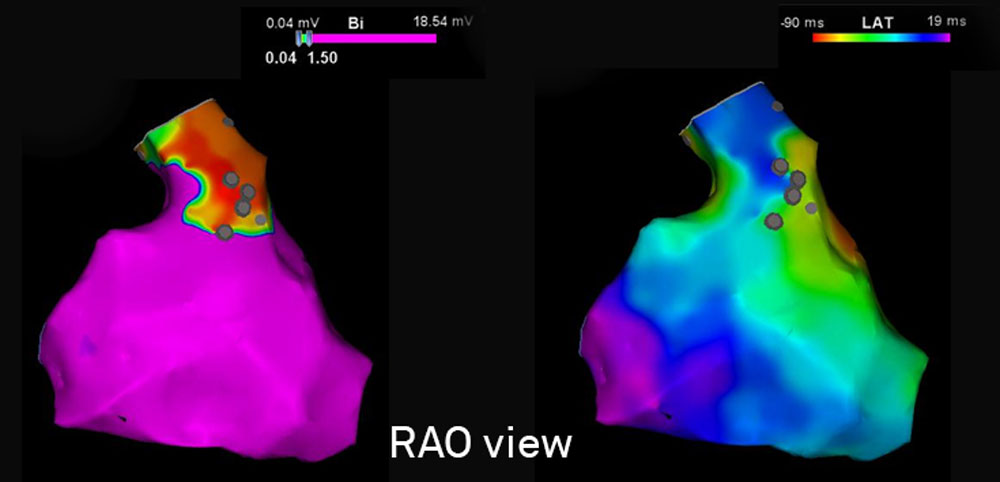
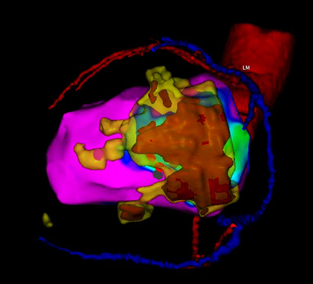
In collaboration with researchers of the technical University Delft and the LKEB our WECAM team has developed a sophisticated program for real-time multimodal image integration. Accurate integration of all imaging-derived data during the mapping and ablation procedure facilitates the procedure and contributes to patient safety and improved outcome. The reversed registration of datasets provides important insights and is key to further develop methods for arrhythmia substrate identification and delineation by imaging.
Projects
Integration of whole heart histology, ex-vivo imaging and arrhythmia substrate mapping
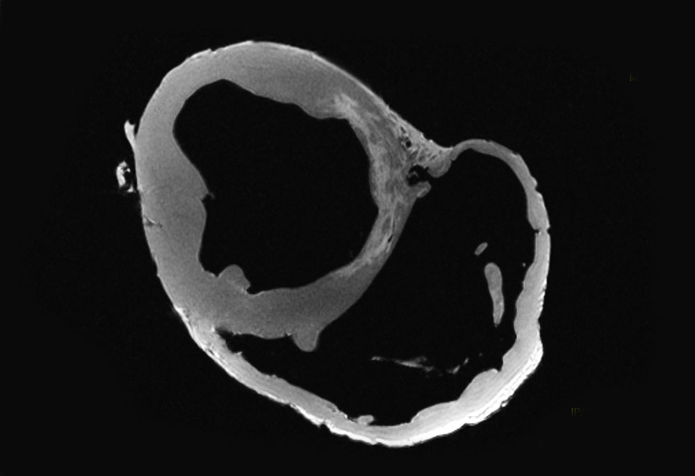
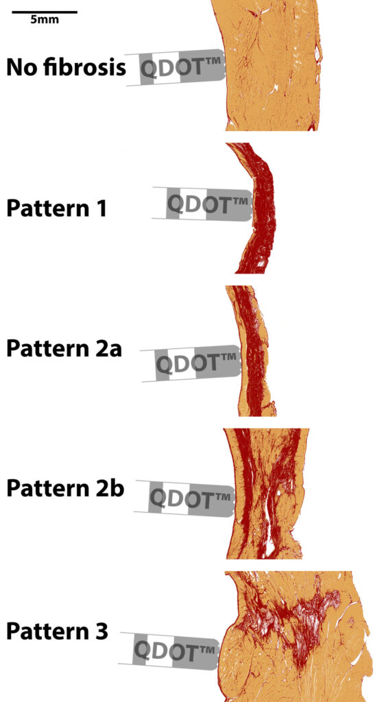
A developed and established workflow for high resolution ex-vivo imaging of pig hearts that underwent an early re-perfusion infarction with accurate integration of in-vivo mapping data and whole heart histology is currently used to validate the performance of novel catheters to delineate scar and arrhythmias substrates.
ACHD substrate visualization
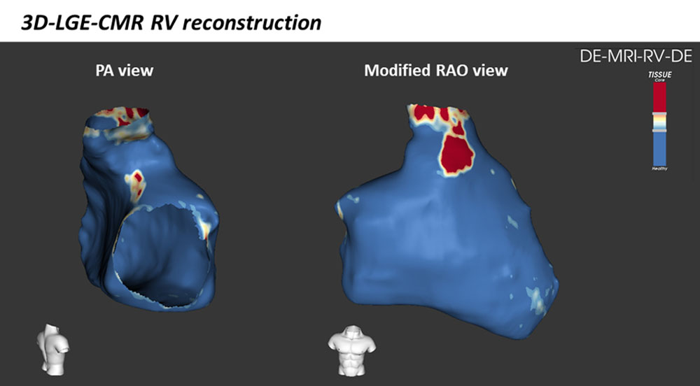
Non-invasive identification of VT substrates in ACHD has not been established. Post-processing of 3D LGE-CMR facilitates accurate reconstruction of morphologically complex areas of the heart such as the RV outflow tract with high-spatial resolution. The performance of this method for non-invasive visualization of arrhythmogenic substrates in ACHD is under investigation.
